An X-ray technique chart is a guide for radiographers, outlining optimal settings for various procedures. It ensures consistency, safety, and quality in radiographic imaging by standardizing parameters like kVp and mAs.
Definition and Purpose
An X-ray technique chart is a standardized document outlining specific exposure settings for radiographic procedures. It defines the optimal kVp, mAs, and exposure time for various anatomical regions. The primary purpose is to ensure consistency, safety, and image quality by standardizing exposure factors. This guide helps radiographers achieve accurate results while minimizing radiation dose. It serves as a quick reference, reducing errors and ensuring reproducible outcomes across different patients and procedures.
Importance in Radiography
X-ray technique charts are essential for ensuring consistent, high-quality radiographic images while minimizing radiation exposure. They provide standardized protocols for various anatomical regions, reducing variability and errors. By optimizing kVp, mAs, and exposure time, these charts enhance image clarity and diagnostic accuracy. They also play a critical role in radiation safety, balancing image quality with patient protection. Standardized techniques streamline workflow, improve efficiency, and ensure reproducible results, making them indispensable in modern radiography practice.
Key Components of an X-Ray Technique Chart
An X-ray technique chart includes kVp, mAs, exposure time, and tube current. These settings optimize image quality and radiation safety for various radiographic examinations.
kVp (Kilovoltage Peak) Settings
kVp (Kilovoltage Peak) settings determine the energy level of the X-ray beam. They regulate the penetration power, ensuring optimal image quality. Higher kVp increases beam energy, improving penetration for denser tissues. Lower kVp is used for softer tissues to avoid overexposure. Proper kVp selection balances contrast and detail, ensuring diagnostic accuracy while minimizing radiation exposure. Technique charts provide standardized kVp values for specific anatomical regions to maintain consistency and safety in radiographic procedures.
mAs (Milliampere-Seconds) Settings
mAs (Milliampere-Seconds) settings control the quantity of X-rays produced, combining milliamperage (current) and exposure time. Higher mAs increases radiation dose and image brightness, while lower mAs reduces dose but may compromise image quality. Technique charts provide standardized mAs values for specific procedures, ensuring optimal balance between diagnostic clarity and patient safety. For example, NIHCS charts specify mAs settings like 2.5 for chest PA and 10 for chest LAT, tailored to anatomical requirements.
Exposure Time and Tube Current
Exposure time and tube current are critical factors in radiography, determining the X-ray beam’s intensity and duration. Technique charts specify these settings to optimize image quality while minimizing radiation exposure. Tube current (mA) regulates the electron flow in the X-ray tube, while exposure time (seconds) controls the beam’s duration. Together, they influence the overall dose (mAs) delivered. For example, NIHCS charts often list exposure times and currents for procedures like chest X-rays, ensuring precise and safe radiographic outcomes tailored to anatomical requirements.

Types of X-Ray Techniques
X-ray techniques are broadly categorized into intraoral and extraoral methods. Intraoral focuses on detailed dental structures, while extraoral captures larger anatomical areas, each requiring specific chart settings for optimal imaging.
Intraoral Radiography Techniques
Intraoral radiography involves placing the X-ray sensor inside the mouth to capture detailed images of dental structures. Common techniques include periapical, bitewing, and occlusal views. Fixed kVp and mA settings are often used, with exposure time as the primary variable. These methods are essential for diagnosing caries, periodontal disease, and other dental pathologies. Technique charts for intraoral radiography are standardized to ensure image quality and patient safety, often referencing guidelines from sources like the NIHCS Exposure Technique Chart Analysis and KODAK Radiography Technique Chart Overview.
Extraoral Radiography Techniques
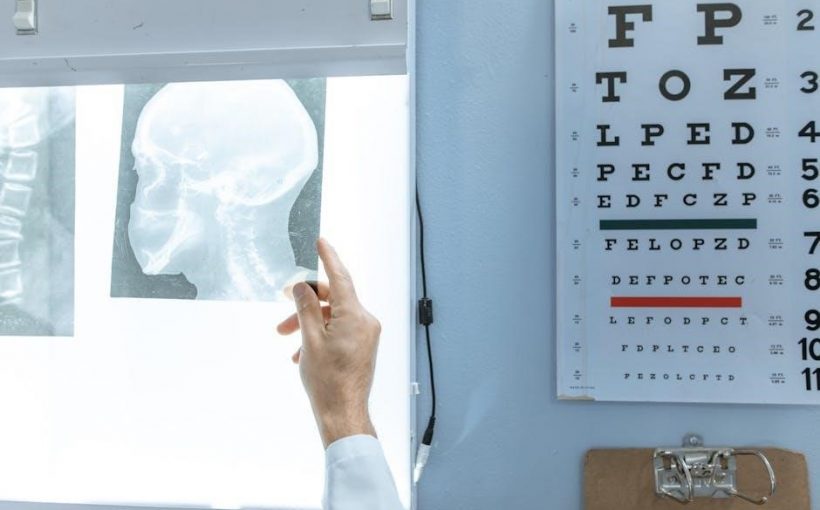
Extraoral radiography involves positioning the X-ray sensor outside the mouth to capture broader anatomical views. Techniques include lateral skull, panoramic, and cephalometric projections, often used for evaluating larger structures like the maxillofacial region. Higher kVp settings are typically employed to penetrate denser bone. These methods are invaluable for diagnosing fractures, developmental anomalies, and pathologies requiring a wider field of view. Technique charts, such as those referenced in the NIHCS Exposure Technique Chart Analysis and KODAK Radiography Technique Chart Overview, guide these procedures to ensure optimal results and patient safety.

Significance of Standardization
Standardization ensures consistency across radiographic procedures, aligns with established protocols like NIHCS and KODAK charts, and enhances patient safety by optimizing radiation exposure.
Uniformity in Radiographic Procedures
Uniformity in radiographic procedures is achieved through standardized technique charts, ensuring consistency in image quality and diagnostic accuracy. These charts, like the NIHCS and KODAK examples, provide predefined settings for various anatomical regions, reducing variability among technicians. By standardizing kVp, mAs, and exposure times, they ensure reliable outcomes across different machines and facilities. This consistency is crucial for accurate diagnostics and Comparability of results. Additionally, uniformity simplifies training and ensures adherence to safety protocols, making radiographic procedures more efficient and reproducible.
Role in Radiation Safety
X-ray technique charts play a vital role in radiation safety by standardizing exposure settings, ensuring minimal radiation dose to patients. They provide optimal kVp, mAs, and exposure times, minimizing unnecessary radiation. By adhering to these guidelines, radiographers reduce the risk of overexposure, protecting both patients and staff. This standardized approach ensures compliance with radiation safety protocols, promoting safer imaging practices while maintaining diagnostic image quality. Technique charts are essential for balancing patient safety with effective radiographic outcomes.
Digital Radiography Considerations
Digital radiography requires technique charts specific to each CR or DR system, ensuring optimal image quality. Updating charts when equipment changes is crucial for consistency and accuracy.
Computed Radiography (CR) Technique Charts
Computed Radiography (CR) technique charts are tailored for systems using phosphor plates. They ensure optimal image quality by standardizing exposure parameters like kVp and mAs for specific anatomical regions. CR charts must account for the unique sensitivity of phosphor plates and the processing algorithms used in CR systems. Properly designed CR charts help minimize radiation exposure while maintaining diagnostic image quality.
Regular updates to CR technique charts are essential when upgrading equipment or changing image receptors. This ensures consistency in radiographic procedures and adherence to safety standards. CR charts are often integrated with digital systems for efficient workflow and dose monitoring.
Digital Radiography (DR) Technique Charts
Digital Radiography (DR) technique charts are customized for specific DR systems and image receptors. They standardize exposure settings like kVp and mAs for various anatomical regions, ensuring optimal image quality. DR charts are dynamic, allowing adjustments based on patient size and anatomy to minimize radiation dose while maintaining diagnostic accuracy.
Unlike CR, DR systems require charts tailored to their unique detectors. Updates are necessary when upgrading hardware or software. DR charts are often integrated with modern imaging systems, enabling streamlined workflows and precise dose monitoring. This ensures consistent, high-quality radiographic outcomes.

Practical Applications
X-ray technique charts are essential in daily radiographic practice, providing standardized settings for various procedures. They guide radiographers in positioning, exposure, and equipment adjustments, ensuring accurate imaging.
Developing a Technique Chart
Developing a technique chart involves tailoring settings for specific X-ray units and anatomical regions. It requires balancing factors like kVp, mAs, and exposure time to optimize image quality while minimizing radiation dose. Calibration of equipment and consideration of patient size variations are critical. The chart should be standardized to ensure consistency across procedures and updated periodically to reflect advancements in technology or changes in equipment. This process ensures radiographers can produce high-quality images safely and efficiently.
Positioning and Exposure Guidelines
Proper positioning and exposure are critical for obtaining high-quality radiographic images. Technique charts provide specific guidelines for anatomical alignment, ensuring accurate representation of body structures. Exposure settings, such as kVp and mAs, are tailored to body part thickness and density. Maintaining the correct tube-to-film distance is essential for image clarity. These guidelines help standardize procedures, reduce retakes, and ensure patient safety by minimizing radiation exposure while optimizing image quality. Consistent adherence to these protocols is vital for reliable diagnostic outcomes.
Portable X-Ray Techniques
Portable X-ray techniques adapt standard radiographic methods for mobile use, ensuring diagnostic accuracy in diverse settings. They are widely used in chest imaging and specialized procedures.
Challenges in Portable Radiography
Portable radiography faces challenges such as scatter radiation exposure, requiring specific technique charts for dose optimization. Maintaining image quality without controlled environments is difficult. Technique charts must be adjusted for varying conditions, ensuring safety and diagnostic accuracy in diverse settings. This complexity highlights the need for standardized protocols to address these unique challenges effectively.
Optimizing Exposure in Portable Settings
Optimizing exposure in portable radiography involves adjusting kVp and mAs to compensate for varying patient sizes and anatomical areas. Technique charts help standardize these adjustments, ensuring diagnostic image quality while minimizing radiation dose. Using grids and collimation reduces scatter radiation, improving contrast. Proper positioning and alignment between the X-ray source, patient, and detector are critical. Regular calibration of portable units and adherence to technique charts are essential for consistent and safe imaging outcomes in non-traditional settings.
Case Studies and Examples
Case studies highlight the application of NIHCS exposure technique charts and KODAK radiography technique charts, demonstrating their role in standardizing exposure settings for consistent imaging results.
NIHCS Exposure Technique Chart Analysis
The NIHCS Exposure Technique Chart provides standardized settings for various radiographic procedures, ensuring optimal image quality and radiation safety. It includes detailed parameters such as kVp, mAs, and exposure times for chest PA, LAT, and portable examinations. These guidelines help maintain consistency across imaging practices, reducing variability and enhancing diagnostic accuracy. The chart also addresses adjustments for different patient sizes and anatomical regions, ensuring tailored approaches for diverse clinical scenarios. Its structured format facilitates efficient workflow and adherence to radiographic best practices.
KODAK Radiography Technique Chart Overview
KODAK’s Radiography Technique Chart offers detailed settings for various radiographic examinations, ensuring consistent image quality. It includes specific kVp and mAs values for chest, cervical spine, and lumbar spine procedures. Designed for both conventional and digital systems, the chart provides standardized protocols, reducing variability in imaging practices. Available in PDF format, it serves as a practical guide for radiographers, promoting efficiency and adherence to diagnostic standards while maintaining patient safety and optimal radiographic outcomes.
Future Trends
Advancements in digital technique charts and integration with modern imaging systems promise enhanced efficiency, improved image quality, and streamlined radiographic workflows in the future.
Advancements in Digital Technique Charts
Digital technique charts are evolving with advancements in computed radiography (CR) and digital radiography (DR). These systems offer enhanced image quality, reduced radiation exposure, and faster processing times. Modern software enables customization of settings for specific anatomical regions, improving diagnostic accuracy. Additionally, digital charts facilitate easier sharing and archiving of radiographic data, streamlining workflows in healthcare facilities. Integration with AI for automated adjustments further optimizes imaging protocols, ensuring consistent and high-quality results across diverse patient populations.
Integration with Modern Imaging Systems
Modern imaging systems seamlessly integrate with digital technique charts, enhancing workflow efficiency and diagnostic accuracy. Systems like PACS (Picture Archiving and Communication Systems) and EHR (Electronic Health Records) allow for centralized access to radiographic data. This integration ensures that technique charts are easily accessible and updatable, reducing errors and improving patient care. Automated data transfer also streamlines the imaging process, enabling radiologists to focus on interpretation rather than manual data entry, thus enhancing overall efficiency and patient outcomes.